As we go about our daily lives, we often take our teeth for granted. However, understanding the structure of a tooth is essential for maintaining good oral health and preventing dental issues. In this comprehensive guide, we will delve into the intricate details of tooth anatomy, from the crown to the roots. So, let’s embark on a journey to unravel the mysteries of our pearly whites.
As we go about our daily lives, we often take our teeth for granted. However, understanding the structure of a tooth is essential for maintaining good oral health and preventing dental issues. In this comprehensive guide, we will delve into the intricate details of tooth anatomy, from the crown to the roots. So, let’s embark on a journey to unravel the mysteries of our pearly whites.
Table of Contents,
The Anatomy of a Tooth: Exploring the Crown and the Roots
A tooth can be divided into two main parts: the crown and the roots. The crown is the visible part of the tooth above the gum line, while the roots are embedded in the jawbone. These two components work together to provide us with the ability to bite, chew, and speak.
The Crown: Unveiling the Outer Layer
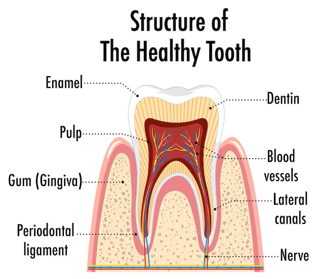
The crown of a tooth is the part that we can see when we smile or open our mouths. It is covered by a protective layer called enamel, which is the hardest substance in the human body. Enamel shields the inner layers of the tooth from damage and decay. Despite its strength, enamel can still be susceptible to wear and tear if not cared for properly.
Beneath the enamel lies another layer called dentin. Dentin makes up the majority of the tooth structure and is softer than enamel. It provides support to the enamel and helps transmit sensory signals to the nerves within the tooth.
The Roots: Anchoring the Tooth
While the crown is the part of the tooth that gets most of the attention, the roots play a crucial role in holding the tooth securely in place. The roots extend into the jawbone and are surrounded by a layer called cementum. Cementum provides a protective covering for the roots and helps anchor them to the bone.
Within the roots, we find the pulp chamber, which houses the delicate pulp tissue. The pulp contains blood vessels, nerves, and connective tissue, and it is vital for the nourishment and sensation of the tooth.
Delving Deeper: Exploring the Layers of a Tooth
A tooth consists of several layers of tissue, each with its own unique function and characteristics. Let’s take a closer look at these layers and how they contribute to the overall structure of the tooth.
Enamel: The Guardian of the Tooth
Enamel is the outermost layer of the tooth and acts as a protective shield against bacteria, acids, and mechanical forces. Composed mainly of minerals, such as hydroxyapatite, enamel is exceptionally hard and durable. However, it is important to note that enamel cannot repair itself once it is damaged, so it is crucial to maintain good oral hygiene practices to preserve its integrity.
Dentin: Providing Support and Sensation
Dentin lies beneath the enamel and forms the bulk of the tooth structure. It is a hard tissue that is slightly softer than enamel but still resilient. Dentin contains tiny tubules that connect to the nerves within the pulp, allowing for the transmission of sensory signals. If the enamel is compromised, the exposed dentin can lead to tooth sensitivity and discomfort.
Pulp: The Heart of the Tooth
Deep within the tooth, we find the pulp, which is a soft tissue comprising blood vessels, nerves, and connective tissue. The pulp is responsible for the development and nourishment of the tooth during its early stages. It also helps to detect sensations such as temperature and pressure. When the pulp becomes infected or inflamed, it can cause severe toothache and may require root canal treatment.
Cementum: Protecting the Roots
Cementum covers the roots of the tooth and provides an anchor for the periodontal ligament, which connects the tooth to the surrounding bone. Cementum is not as hard as enamel or dentin but serves an essential role in protecting the sensitive root surfaces. It helps to maintain the stability of the tooth within the jawbone.
Periodontal Ligament: The Tooth’s Suspension System
The periodontal ligament surrounds the roots of the tooth and attaches them to the jawbone. The ligament is made up of collagen fibers that act as a shock absorber, allowing the tooth to withstand the forces of biting and chewing. It also provides a cushioning effect, preventing damage to the tooth and the surrounding bone.
Gingivae: Safeguarding the Tooth-Gum Interface
The gingivae, commonly known as the gums, are the soft tissues that surround the teeth and provide a protective barrier against bacteria and debris. Healthy gums are crucial for maintaining good oral hygiene and preventing gum disease. Regular brushing, flossing, and professional dental cleanings can help keep the gums healthy and free from infection.
Bone: The Foundation of Oral Health
The jawbone provides the structural support for the teeth, holding them in place within the oral cavity. Healthy bone density is essential for maintaining the stability of the teeth and preserving overall oral health. Periodontal disease, poor oral hygiene, and certain medical conditions can compromise the jawbone, leading to tooth loss and other dental issues.
Types of Teeth: Function and Characteristics
Now that we have explored the different layers of a tooth, let’s shift our focus to the types of teeth and their unique functions within the oral cavity.
Incisors: The Cutting Edge
Incisors are the front teeth located in the center of the mouth. They have a thin, sharp edge that is ideal for cutting and biting into food. Incisors play a crucial role in the initial stages of digestion, as they help break down food into smaller, more manageable pieces.
Canines: The Mighty Fangs
Canine teeth, also known as cuspids, are situated on either side of the incisors. They have a pointed shape, resembling the fangs of certain animals. Canines are designed for gripping and tearing food, allowing for better control and manipulation during chewing.
Premolars: The Transition Teeth
Premolars, also called bicuspids, are located between the canines and the molars. They have a flatter surface with one or two cusps, making them versatile for both cutting and grinding food. Premolars assist in the initial stages of breaking down food before it reaches the molars for further processing.
Molars: The Power Grinders
Molars are the back teeth that have a larger, flatter surface area with multiple cusps. They are responsible for crushing and grinding food into smaller particles, making it easier to swallow and initiate the digestion process. The molars are essential for efficient chewing and ensuring thorough breakdown of food before it enters the digestive system.
Wisdom Teeth: The Late Bloomers
Wisdom teeth, also known as third molars, are the last set of teeth to erupt, typically during late adolescence or early adulthood. However, not everyone develops wisdom teeth, and in some cases, they may be impacted or require extraction due to a lack of space in the jaw. Wisdom teeth have limited functional significance in modern humans, but they can sometimes cause issues such as overcrowding or impaction.
Maintaining Optimal Oral Health: Tips and Best Practices
Now that we have explored the intricate details of tooth structure and the different types of teeth, let’s discuss some essential tips and best practices for maintaining optimal oral health:
- Brush your teeth at least twice a day with a fluoride toothpaste to remove plaque and bacteria.
- Floss daily to clean between the teeth and along the gumline, where a toothbrush cannot reach.
- Limit your consumption of sugary and acidic foods and beverages, as they can contribute to tooth decay.
- Visit your dentist regularly for professional cleanings and check-ups to detect and address any dental issues early on.
- Wear a mouthguard during sports activities to protect your teeth from injury.
- Avoid tobacco use, as it can lead to gum disease, tooth discoloration, and oral cancer.
- Practice good oral hygiene habits from a young age and encourage children to develop healthy dental care routines.
By following these tips and incorporating them into your daily routine, you can maintain strong and healthy teeth for a lifetime.
In conclusion, understanding the structure of a tooth is crucial for maintaining optimal oral health. From the crown to the roots, each component plays a vital role in the functioning and well-being of our teeth. By practicing good oral hygiene, visiting your dentist regularly, and staying informed about tooth structure and care, you can ensure a confident and radiant smile for years to come.
Remember, your teeth are not just tools for chewing; they are an integral part of your overall well-being. Take care of them, and they will take care of you.
Additional Information:
If you have any concerns or questions about your oral health, don’t hesitate to schedule an appointment with us. We are here to provide personalized care and support, ensuring that you receive the highest quality treatment and guidance on your oral health journey.
Important Links
https://my.clevelandclinic.org/health/body/24655-teeth
https://drkamihoss.com/tooth-structure-101-breaking-down-structure/
https://www.kenhub.com/en/library/anatomy/anatomy-of-the-tooth
MedicAL DISCLAIMER
The information available on the Website is for general health information only and is not intended to be a substitute for professional medical advice, diagnosis or treatment. You should not rely exclusively on information provided on the Website for your health needs. All specific medical questions should be presented to your own health care provider and you should seek medical advice regarding and before making any changes related to your health.